

The implant behind the muscle at a glance
- PositionUnder the pectoralis major muscle (upper part of the implant)
- AdvantageImplant disguised in the cleavage
- Capsular contractureLess frequent than in front of the muscle
- DrawbackMovement when the muscle contracts
- Most used variantDual plane
- Price€5,000 to €10,000
In a subpectoral breast augmentation, known as “behind the muscle”, the upper and inner parts of the implant are placed under the pectoralis major muscle, which disguises it in the cleavage. This position limits rippling and the feeling of an implant under the skin, makes capsular contracture less frequent and reduces long-term sagging of the breast. On the other hand, recovery is more painful and the implant moves when the muscle contracts. Dr Médard de Chardon most often uses a variant, the dual plane, which keeps these advantages while limiting the drawbacks.
Price: breast augmentation €5,000 to €10,000, implants included, on a written quote after a consultation (€100).
Where is the implant placed?
Despite its name, the implant is only partly behind the muscle: only its upper and inner parts, in the cleavage area, are covered by the pectoralis major muscle. This extra coverage is what makes the position worthwhile in slim patients.
In front of the muscle, behind the muscle or dual plane: what is the difference?
| In front of the muscle (prepectoral) | Behind the muscle (classic subpectoral) | Dual plane | |
|---|---|---|---|
| Implant position | Under the breast gland, in front of the muscle | Upper and inner parts under the muscle | Upper part under the muscle, lower part under the gland |
| Implant disguised in the cleavage | Less well in a slim patient | Yes | Yes |
| Capsular contracture | More frequent | Less frequent | Less frequent |
| Movement when the muscle contracts | No, the implant moves with the breast | Yes | Reduced |
| Recovery | Less painful | More painful, later return to activity | Drawbacks of the subpectoral position reduced |
| Long-term sagging of the breast | More frequent | Less frequent | Less frequent |
| Best indication | Sufficient tissue, slight ptosis, small-volume implant | Insufficient coverage: slim patient, juvenile breast | Most augmentations |
Behind the muscle: the top and the inner side of the implant are covered by the pectoralis major muscle.
- 1Implant
- 2Pectoralis major muscle
- 3Breast gland
- 4Ribs
What are the advantages of the position behind the muscle?
- A better-disguised implant. The upper and inner parts of the implant are covered by the muscle: this prevents the implant from being felt under the skin (palpability) and rippling from appearing in the cleavage.
- Less capsular contracture than in front of the muscle, thanks to the constant massaging effect of the muscle and probably to a better blood supply to the pocket.
- No loss of strength: the function of the pectoralis major muscle is not altered.
- Less sagging of the breast (ptosis) in the long term than in front of the muscle.
What are its drawbacks?
- Recovery is more painful and the return to normal activity is later than with an implant in front of the muscle.
- The implant moves when the muscle contracts: the breast shifts slightly with certain arm movements.
- The risk of the implant shifting (malposition) is thought to be higher than in front of the muscle if the pocket is not made perfectly.
These drawbacks are less marked with the dual plane. Choosing a qualified, experienced and reputable surgeon reduces these risks, and can even eliminate some of them.
Who is the position behind the muscle suited to?
According to Dr Médard de Chardon’s research (2008 thesis), the best indication is insufficient coverage of the implant: a slim patient, little breast tissue, thin skin and tissues, a narrow breast base. The best example is the juvenile breast, small and firm.
When should it be avoided? The classic subpectoral pocket is not suited to a small, sagging breast with poor-quality skin (hypotrophy with ptosis): the skin and breast tissue drape poorly over the implant, with a risk of the implant shifting and of a double contour of the breast (the breast appears to slide down in front of the implant). The dual plane is then more suitable.
The dual plane: the most widely used position
In the dual plane, the upper part of the implant is behind the muscle and the lower part in front of it, directly under the breast gland. To achieve this, the surgeon releases the lower attachments of the pectoralis major muscle, which moves up and then covers only the upper part of the implant. The dual plane keeps the advantages of the position behind the muscle (implant disguised in the cleavage, less capsular contracture) and those of the position in front of the muscle (the skin and breast tissue drape well over the implant). It is the position Dr Médard de Chardon chooses in most cases.
Dual plane type I, II or III
The type indicates how far the gland is separated from the muscle, and therefore how far up the edge of the muscle rides over the implant:
- Type I: only the lower attachments of the muscle, along the fold under the breast, are released. For a breast without ptosis, with firm breast tissue.
- Type II: in addition, the gland is separated from the muscle up to the lower edge of the areola. For mobile breast tissue or a slightly sagging breast.
- Type III: the separation goes up to the upper edge of the areola. For a deflated or sagging breast (glandular ptosis).
The higher the type, the better the breast tissue drapes over the implant. The type is chosen in consultation, based on examination of the breast.
Type I: only the lower attachments of the muscle, along the fold under the breast, are released. For a breast without ptosis, with firm breast tissue.
Dr Médard de Chardon’s experience
- MD thesis on breast augmentation with implants, defended at the Nice Faculty of Medicine in 2008: the indications above are drawn from it.
- More than 3,000 breast implants placed; practising in Cannes since 2011.
- Qualified plastic surgeon in plastic, reconstructive and aesthetic surgery, former Chef de Clinique (university hospital senior registrar), Nice Faculty of Medicine; member of SOFCEP (French Society of Aesthetic Plastic Surgery), SOFCPRE (French Society of Plastic, Reconstructive and Aesthetic Surgery) and ISAPS (International Society of Aesthetic Plastic Surgery).
How does breast augmentation work?
The position of the implant (in front of the muscle, behind the muscle or dual plane), its shape, its volume and the scar are chosen in consultation, after trial sizers and a 3D simulation. The operation lasts about 1 hour, as day surgery or with a one-night stay at Clinique Oxford in Cannes; time off work is 7 to 10 days. The process, recovery and risks are set out on the breast augmentation page. See also: round implants, anatomical implants.
Before and after photos
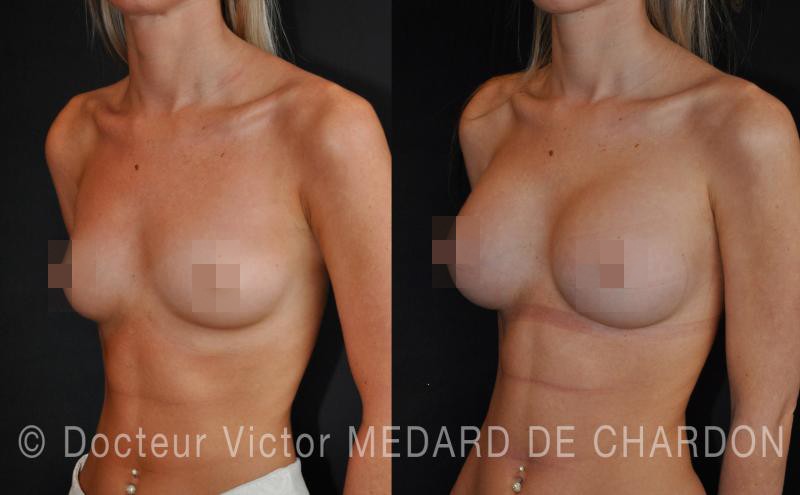
Breast augmentation with implants through an inframammary incision, dual plane, 320 cc
Breast hypoplasia on a narrow chest, slim patient
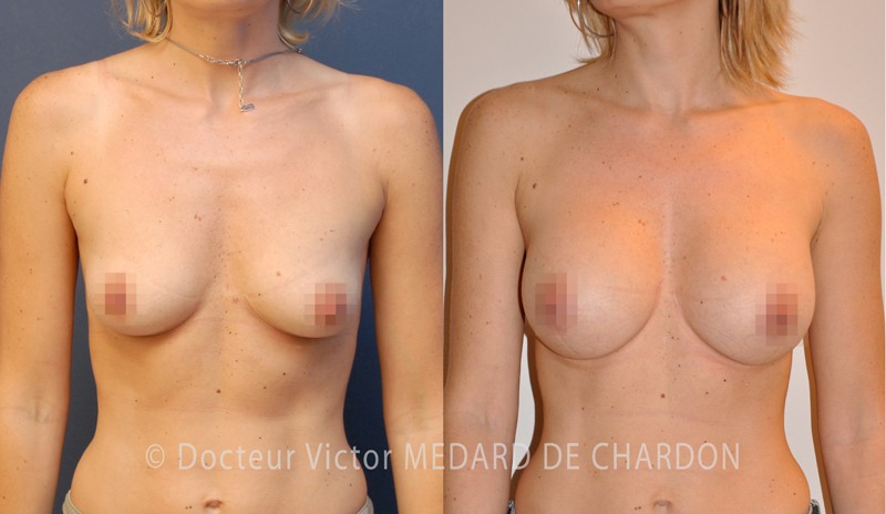
Breast augmentation with 275 cc moderate-profile round breast implants, dual plane II, through an areolar incision
Breast augmentation with breast implants
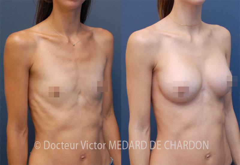
340 cc anatomical breast implants, inframammary incision, dual plane I
Breast augmentation with breast implants
See all before and after breast augmentation photos
Frequently asked questions about the implant behind the muscle
Breast implant behind or in front of the muscle: which should you choose?
It all depends on the thickness of the tissues covering the implant. In a slim patient, the position behind the muscle disguises the implant better in the cleavage; when the tissues are sufficient and the implant small, the position in front of the muscle is possible. Dr Médard de Chardon most often uses the dual plane, which combines the advantages of both.
What is the dual plane in breast augmentation?
It is a variant of the position behind the muscle: the upper part of the implant is covered by the pectoralis major muscle, and the lower part is placed directly under the breast gland. The implant remains disguised in the cleavage and the skin drapes well over it.
What do dual plane I, II and III mean for a breast implant?
The number indicates how far the gland is separated from the muscle: up to the fold under the breast (I), up to the bottom of the areola (II) or up to the top of the areola (III). The more sagging or deflated the breast, the higher the type chosen.
Is placing a breast implant behind the muscle more painful?
Yes, recovery is more painful and the return to normal activity is later than with an implant in front of the muscle. These drawbacks are reduced with the dual plane.
Does the breast implant move when you contract the pectoral muscle?
Yes, in the position behind the muscle, the implant and the breast shift slightly when the pectoralis major muscle contracts. This movement is reduced with the dual plane.
Does the pectoral muscle lose strength with an implant placed behind it?
No. An implant placed behind the muscle does not alter the function of the pectoralis major muscle.
Does placing the breast implant behind the muscle reduce the risk of capsular contracture?
Yes, capsular contracture is less frequent than in front of the muscle, thanks to the constant massage exerted by the muscle and probably to the better blood supply to the pocket.
References
This information is drawn from Dr Victor Médard de Chardon’s MD thesis on breast augmentation with implants (Nice Faculty of Medicine, 2008).
- Hidalgo DA. Breast augmentation: choosing the optimal incision, implant, and pocket plane. Plast Reconstr Surg 2000; 105: 2202-2216.
- Vazquez B, Given KS, Houston GC. Breast augmentation: a review of subglandular and submuscular implantation. Aesthetic Plast Surg 1987; 11: 101-105.
- Camirand A, Doucet J. Breast augmentation: teaching our patients how compression can help prevent capsular contracture. Aesthetic Plast Surg 2000; 24: 221-226.
- Banbury J, Yetman R, Lucas A, et al. Prospective analysis of the outcome of subpectoral breast augmentation: sensory changes, muscle function, and body image. Plast Reconstr Surg 2004; 113: 701-707.
- Heden P, Jernbeck J, Hober M. Breast augmentation with anatomical cohesive gel implants: the world’s largest current experience. Clin Plast Surg 2001; 28: 531-552.
- Araco A, Gravante G, Araco F, et al. A retrospective analysis of 3,000 primary aesthetic breast augmentations: postoperative complications and associated factors. Aesthetic Plast Surg 2007; 31: 532-539.